The pictures below are helpful in understanding what's happening inside Jordan. The first picture shows the trachea and the left and right bronchi. When they suction Jordan out, they are suctioning out the trachea. Since his lungs are closed due to inflammation, air from the ventilator only enters the bronchi.
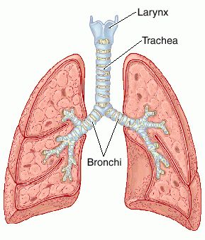 |
| Image of the Trachea from thefreedictionary.com |
Below is an image of a healthy baby's chest x-ray. You can see that the left and right sides of the body appear black and you can easily count the ribs. Also, the heart on the lower left-hand side of the chest.
 |
| Healthy Baby's Chest X-ray from sciencephoto.com |
In contrast, in Jordan's current chest x-ray, both sides of the chest appear white where the lungs are closed. The bottom photo is one of the best we found that shows what CDH looks like under x-ray. Jordan has had his repair, so the major difference with him now is that the bowel is not up in his chest.
 |
| Chest X-ray of a Baby with CDH from cdhgenetics.com |
No comments:
Post a Comment